How Vets Evaluate Lameness in Horses

How Vets Evaluate Lameness in Horses

Whinny’s Wisdoms

Springhill Equine Veterinary Clinic

Hi everyone, Whinny here! In the previous installment of my Lameness Series, we talked about what you might notice if your horse is lame and needs a visit with one of my Springhill vets. This week, we’re going to talk about what happens once you make that appointment! There are several parts to a lameness exam that help my vet figure out the problem, and you might see her doing some or all of these things to check out your horse. Some types of lameness are more obvious and won’t require all the steps, but for a more subtle lameness, each of them can be a piece in the puzzle to find the source of the problem.

Springhill Equine Veterinary Clinic

Physical Exam

My vet will do an exam to look and feel for abnormalities in the horse’s body. She will look at his conformation and muscle symmetry. She will check out his posture to see if he stands squarely, since abnormal stance can give clues to the areas of discomfort. His hooves and shoes will also be evaluated carefully – I’m sure you have heard the saying “No hoof, no horse” – the angles and health of the hooves are critically important to soundness. My vet will feel your horse’s limbs for heat or swelling and evaluate his tendons for thickening or pain. She’ll feel the limb pulses to look for areas of inflammation. The joints will be palpated for swelling and taken through their range of motion. The exam will often include an evaluation of the back muscles for tension or sensitivity, especially with a hind end lameness.
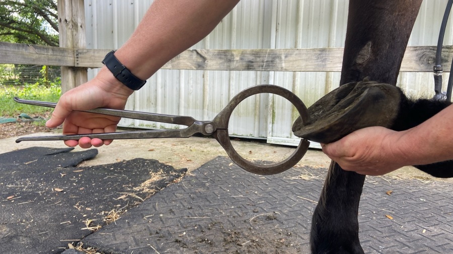
 Hoof Testers

You may see my vets use a big metal instrument to squeeze your horse’s hoof. That instrument is called a hoof tester and it checks for sensitivity when pressure is applied to certain areas of the hoof. My vet’s knowledge of the anatomy inside the hoof helps her to determine what pain in a certain spot means. Sensitivity in one area may indicate laminitis, whereas another spot may signify a problem with the navicular bone. Hooftesters can also be used to locate the position of a hoof abscess so my vet can open it up to drain.

Flexion Tests

You may see my vet holding up your horse’s leg for 30 or 45 seconds and then asking him to immediately trot off – that’s a flexion test. The purpose of a flexion test is to accentuate pain that may be coming from a joint, in order to localize the part of the leg that is bothering your horse or to look for a subtle problem that isn’t immediately apparent. Specific joints are flexed in turn to check the response to that area. If your horse has an arthritic hock, for example, flexing the hock may make him trot off more lame than he was without the flexion. That can help my vet determine the part of the leg that needs treatment. Flexion tests aren’t always a black or white answer, but they can be a useful puzzle piece in some cases. My vet uses her experience to know the appropriate position, time, and pressure for a flexion test, since it’s possible to get an inaccurate assessment if you flex the joint too hard or for too long. It’s also useful for her to flex the same joint on both the left and right sides to compare how the horse responds.

Springhill Equine Veterinary Clinic

Nerve or Joint Blocks

Have you ever gone to the dentist and had a shot to make your tooth numb for a filling? That’s basically the same thing as a nerve block my vet may use during a lameness exam. When my vet “blocks out” an area on your horse’s leg, she is temporarily numbing it to see if that region is the source of the pain. If the correct spot is numbed, your horse won’t look lame anymore since he won’t feel the pain. Unless there is an obvious abnormal finding on her physical exam, my vet will inject a numbing agent into specific anatomical areas until she finds the one that takes away the lameness. Nerve blocks are an injection to directly numb a nerve and the area it supplies feeling to, while joint blocks will inject the numbing agent right into a joint, which is a sterile procedure. Blocks only lasts a couple of hours though, so don’t confuse them for a permanent treatment, they are just a way of finding where the problem is so it can receive the appropriate therapy.

Imaging

Once my vet has determined which leg your horse is sore on and which part of the leg is the problem, she will often recommend imaging to get a look at what’s going on inside. This is most often an X-ray (radiograph) or ultrasound. Generally speaking, X-rays look at bone and ultrasound looks at soft tissues such as tendons.

Occasionally, advanced imaging such as CT or MRI is needed, but the majority of cases can be diagnosed with the imaging equipment my vet carries in her truck. Once a specific diagnosis is made, she can recommend the best treatment to get your horse sound and back doing those things you want to do with him!

If your horse has something going on, make sure to give my team here at Springhill Equine a call to schedule a lameness evaluation. They are amazing at what they do!

Until next week,

~Whinny

P.S. Are you missing out? In addition to my amazing blog, this website also has the biggest equine podcast in the world, a huge video library filled with how-to’s and seminars, a series of best-selling books, and direct access to some of the best equine veterinarians on the planet! I’m just a small (pun intended!) part of a fabulous team of equine professionals here at Springhill, and I want to make sure you are taking advantage of everything we have to offer. And most of it is free! So take a minute to explore the rest of the website while you’re here. I promise you’ll find something you can use to make the world a better place for your horse.

Whinny’s Wisdoms is the official blog of Whinny the Clinic Mouse at Springhill Equine Veterinary Clinic in Newberry, Florida. If you liked this blog, please subscribe below, and share it with your friends on social media! For more information, please call us at (352) 472-1620, visit our website at SpringhillEquine.com, or follow us on Facebook!

Subscribe to Whinny's Wisdoms

Enter your email address to subscribe to this blog and receive notifications of new posts by email.

More Adventures of the Horse Doctor's Husband
What Does Lameness Look Like?

What Does Lameness Look Like?

Whinny’s Wisdoms

Springhill Equine Veterinary Clinic

Hi everyone, Whinny here! It’s starting to cool off a teeny bit here in Florida, and a lot of you are gearing up for the riding season! We don’t want anything to get in the way of the fun stuff you’re planning with your horse this Autumn. My Springhill Equine vets love to keep your horse feeling good and performing up to his potential, no matter whether you’re a show jumper, reiner, trail rider, or anything in between!

Lameness evaluation and treatment is one of the most common things they work on. But the first step is for you to recognize the start of an issue at home, so let’s talk about what to look for. Some lameness is really obvious, like if your horse is limping badly on one foot, but a lot of the time it’s much more subtle! You may just notice a change in his performance, such as trouble making tight turns around the barrel, difficulty picking up one canter lead, or acting cranky out on the trails. Once you notice something is amiss, here’s what my vets will look for when they’re evaluating your horse move.

What does lameness look like?

If you drive by my clinic, you will often see horses being trotted up and down the grass outside the clinic while my vets watch them. This is the Springhill technicians’ favorite part of the day. They just love the healthy exercise, especially at noon in July and August! 

So what does it look like when a horse is lame? It can be an obvious limp in a forelimb lameness. Your horse may not want to bear weight on the leg or may have a pronounced “head bob” when he walks or trots. For a forelimb lameness, think “down on sound” – meaning the head bobs downwards when the sound (non-lame) leg hits the ground. So, if the head bobs down when the left front foot hits the ground, the lameness is on the right front leg. For a mild lameness, there may not be a super obvious head bob, especially when he’s moving on a straight line. My vets will also listen to the sound of his footfalls – a lame horse will land softer on the foot that hurts, as he shifts his weight to avoid pain. If both forelimbs hurt equally, you might not see a head bob, but your horse could look stiff when he’s moving, or rock his weight back onto his hind end to take weight off the front.

Springhill Equine Veterinary Clinic

Hindlimb lameness is evaluated differently than forelimb lameness. It may look like a “hip hike”, toe dragging, or a shortened stride on the lame hindlimb. It can also just manifest as problems picking up or keeping the canter leads, lack of impulsion, or even “crow hopping” or bucking.  Don’t worry if you aren’t sure, hindlimb lameness can be tricky to see – that’s what my vets are for. If you’re feeling like something doesn’t seem right, just call us!

Some lameness is more subtle than others. If your horse is quite sore, my vets may just need to see him walk and may not need him to trot. For most lameness cases though, the horse is evaluated at the trot, because the symmetrical nature of the normal trot gait makes abnormalities more apparent. Sometimes my vet will ask to see a horse canter to check out how the hind end moves. She’ll want to see the horse move on a straight line and may want to see him moving in a circle (such as on a longe line) to see how positioning his limbs on the inside or outside of the circle will affect his lameness. Some lameness is only noticeable when the horse is turning. The ground surface makes a difference too – your horse may look fine on grass or soft sand but be sore when the ground is harder. That’s typical of a hoof problem or arthritis. A soft tissue injury like tendinitis may be more painful on deep, soft footing. So my vet may ask her tech to jog your horse on an asphalt surface instead of the grass to check out the difference.

Here’s one thing I want you to understand – horses don’t lie about lameness. Their brains don’t work that way. Your horse isn’t “faking” a lameness when you ride him just because he doesn’t look as lame when you see him cantering in the field. He isn’t capable of that. Some things, like the additional weight of carrying a rider, or the specific motions he is asked to do under saddle, just make a lameness more apparent. So, if you notice lameness when you ride, just schedule a lameness exam with my vets instead of thinking your horse is being tricksy.

In my next blog on the lameness exam, we’ll talk about the steps my Springhill vets take to determine what’s hurting your horse, and how to help him!

Until next week,

~Whinny

P.S. Did you know the humans here at Springhill Equine have the biggest, most popular equine veterinary podcast in the world? It’s called Straight from the Horse Doctor’s Mouth. Each episode (and there are over 130 and counting) covers a horse-health topic and is directed at horse owners so they can learn how to take the best care of their horses. You can check it out right over on the Podcast Page of my website, or subscribe on Spotify, Apple Podcasts, or wherever you get your podcasts.

Whinny’s Wisdoms is the official blog of Whinny the Clinic Mouse at Springhill Equine Veterinary Clinic in Newberry, Florida. If you liked this blog, please subscribe below, and share it with your friends on social media! For more information, please call us at (352) 472-1620, visit our website at SpringhillEquine.com, or follow us on Facebook!

Subscribe to Whinny's Wisdoms

Enter your email address to subscribe to this blog and receive notifications of new posts by email.

More Adventures of the Horse Doctor's Husband
Subtle Lameness

Subtle Lameness

Tuesdays with Tony

Ah, the subtle lameness. This is often a source of medical mystery for my Docs. For me, it’s like a good Netflix drama. Is the problem even what you think it is? One minute you’re sure it’s the left rear leg, but it turns out it’s really the neck, and sometimes it isn’t even a lameness! It’s a neurologic issue, or even a muscle disorder. I tell you, it’s high suspense around here! How do my Docs even begin to unravel the mystery? Read on while I drop some cat wisdom on you.

Start at the Beginning

Everyone always wants lameness exams to start with trotting, or riding, or something where they can show off the lame part. My Docs never start here. It’s even more important with the vague lameness. They start by talking with you about what’s going on. The story is often long, complicated, and can have some subtle plot points. 

It may be that it all started with a day where Flicka didn’t feel quite right. From there, some days have been good, maybe some bad. There could be a buck when turning left and picking up the canter. There could be a skip when coming down to a trot from the canter. All those little clues help my Docs begin to formulate an idea. That idea is just that, an idea. It’s likely to change as more information is added, but it’s the starting point of what’s really going on. Much like that Netflix show, there may be some false clues in here, but without exploring them all, my Docs often can’t know what’s real and what’s not. 

Hands On

We’re still not going to run the horse around. The next step is a full cat scan… er, human scan. I’m generally very involved from a supervisory standpoint for this part. My Docs will poke, prod, flex, extend, and turn all your horse’s parts while standing still. 

Each Doc goes through this a little differently, but the general idea is to get their hands on every part of your horse, and to move those parts through their normal range of motion while standing still. This is often very telling about where horses are stiff, sore, or generally reactive. Excellent clues are gained from this section of the exam. Those ideas from earlier are expanded upon, modified, and sometimes changed all together. 

Finally, We Move

Once there has been time spent on history, and hands laid upon the horse, then we head out to see how they move. Generally, my Docs start on grass footing. They watch the horse walk away from them, towards them, and from the side. They will often watch, and then video. Then they do the same at the trot. Depending on how that goes, next may come flexions, or heading to a hard surface to see what happens there, or some circles in both directions. 

Springhill Equine Veterinary Clinic

Different problems show up in different places. Some things don’t like hard surfaces, some don’t like soft, some don’t like to be on the inside of a circle, and some object strongly to being on the outside of the circle. Especially with a subtle lameness, my Docs are going to run through a lot of scenarios. They are also going to video a lot of those scenarios and watch them in slow motion. Slow motion can really highlight an area that isn’t moving normally. Turns out those phones are good for something more than Facebook! 

A Cautionary Tale of Flexions

Flexions are a let’s stress this area and see what happens kind of test. They are far from perfect, but they can sometimes point to an area that hurts when it’s bent, held, and then asked to move. However, flexions can lie in all sorts of ways. Occasionally, there’s a well-performing horse with no issues that trots off like it has a broken leg when flexed. More often there are lame horses that trot off exactly the same when flexed. Flexions add information to the mystery; they don’t solve it!

Springhill Equine Veterinary Clinic

Even More Moving

With a subtle lameness, my Docs will often have you ask the horse to perform their normal job. My Docs will watch the horse go through their paces. They nearly always video this part, especially if faster gaits or quick turns are involved. Even my Docs are only human, and videos help them see the subtle changes going on. For many of these not-so-obvious lamenesses, it takes until this step to really fully complete that inkling of an idea that happened way back at talking to you about history. 

Next Steps

Depending on all the previous steps, the next step can be a variety of things: blocking, radiographs, or ultrasound. Blocking involves numbing an area to see if the problem stops. There are obvious issues with this system. Sometimes you have what we call an unblockable area. Many back and pelvis issues fall under these categories. The other issue is some horses really change the way they move when some part of their body is numb. Last week we had one that held his leg up in the air because his foot was blocked. Very, very difficult to continue a lameness like that! There was nothing to do but wait for the block to wear off and try a different plan. Ah horses. 

Some areas my Docs image before they block if they are suspicious. Proximal suspensories often fall in this category. To block them, you have to put liquid where the ultrasound then looks, which makes it look like you have a really, really bad suspensory issue when it might just be your block. Some areas, like backs, don’t always respond great to blocks so x-rays or ultrasound may be the next step. 

Next, Next Steps

After narrowing down their ideas to a primary problem, my Docs will work on a plan to address that primary issue. This may involve injections, rehab therapy, spinal manipulation, acupuncture, a change in tack, or any of a number of things. Often there is a combo pack of things that need to be done to help these horses. Rarely is it a let me give your horse this magic injection (or pill) and all will be fixed! 

Subtle lamenesses can be frustrating. Making sure you have reasonable expectations for the process, and knowing the results often require a lot of work can help everyone arrive at the best answer for the horse. Unlike Law & Order, the perpetrator isn’t always found in 47 minutes. Sometimes it can be an 8-episode series, and there’s no binge watching allowed. We hope for a 2-3 episode max series to solve the mystery, but horses have their own ideas about what’s entertainment.

One thing I didn’t mention was the rider. An unbalanced rider can make it difficult for the horse to do all the things. That’s a whole different blog, and may require some professional help for the rider (physical therapist to figure out your body, mental therapist to help you accept that you’re not perfect, etc). My humans just released a podcast episode about that very topic called Unbalanced Riders, so make sure you’re subscribed to the show, or head over to the Podcast Page of my website to listen in!

Until next week,

~Tony

P.S. Be a good human and scroll down to the purple box and subscribe to my blog. Don’t rely on Facebook to let you know when a new one is out. My subscribers get it right in their email inbox, and a day earlier than everyone else! Go ahead, just scroll down a little further. Good human. You can do it!

Tuesdays with Tony is the official blog of Tony the Clinic Cat at Springhill Equine Veterinary Clinic in Newberry, Florida. If you liked this blog, please subscribe below, and share it with your friends on social media! For more information, please call us at (352) 472-1620, visit our website at SpringhillEquine.com, or follow us on Facebook!

Subscribe to Whinny's Wisdoms

Enter your email address to subscribe to this blog and receive notifications of new posts by email.

More Adventures of the Horse Doctor's Husband
To Lunge or Not To Lunge

To Lunge or Not To Lunge

Tuesdays with Tony

Those crazy sticks that horses walk around on. It’s not a great design. Then you humans do crazy things like lunging. I mean, I get it. Horses worry about big cats, like me, attacking them out of nowhere, especially if they’re in a new environment like a horse show. A great way to get them to worry less about make-believe monsters is to make sure they’re a bit on the tired side. Lunging has become the preferred method for this. Let’s talk about why that’s not such a great idea for the long term soundness of your best friend. 


Circles. All the circles


Let’s begin with the obvious. Lunging involves horses going around in a circle on a long lead rope. Most of those lead ropes are in the 30 foot range. For a human, this would be like running around on about a 10 foot diameter circle. Try it for a while, and see how it feels. I’ll give you the answer: if you are going to the left, after about 5 minutes the inside of your right foot and the outside of your left foot are going to get sore. Keep going and your knees are going to get sore. Throw in a buck, and a rear while someone pulls on your head just to see how that goes. I think you probably get the idea. 

Lunging is really, really hard on the joints. It’s even harder on the joints when your horse is wild, and doing the gallop whilst leaning at a 45 degree angle to the ground, oh and adding some airs above the ground work. You can see where this could be really, really hard on those sticks they run around on. Add in a foot that’s off balance, and you have a real recipe for disaster. Shameless plug for my many foot blogs. It’s a very important topic, and that’s why I talk about it A LOT.


Options other than circles


I get it. I’ve seen what those crazy horses do when they decide the world is a scary place. I certainly wouldn’t want to ride them when they’re like that! This is a good time to evaluate options before it’s time to tame the wild beast. I hear trainers talk about the importance of transitions all the time. Asking your horse to walk, trot, walk, canter a few steps, walk, while on the lunge line is a great way to get their brain thinking. 

Teach them this before the horse show. Don’t just stand in one place while your horse runs around you. Move around. This will make the circle bigger, which will put less stress on your horse’s joints. If your horse is a real wild one at shows, you may want to consider teaching them how to lunge in a surcingle, and side reins. This will give you a whole new level of control. A surcingle and side reins also help your horse work while lunging, instead of running around in circles. 

Teaching Zen


My favorite option is the ‘not lunging’ option. There are lots of ways to do this. Finding the way that works best for you is key. My favorite is groundwork. There are approximately 8,432 natural horsemanship videos on YouTube. It’s a good place to start getting an understanding of what good groundwork can do for you, and your horse. I myself didn’t understand this whole groundwork thing for a very long time, but I now know it’s all about teaching your horse to stay calm when you say it’s ok. Yes, there can be lots of circles involved in that process, but these can be done at home, and in a controlled fashion. Once your horse understands the principles, there are very few circles, and you can “catch” the brain pretty quickly in even the craziest of horse show worlds. Hand walking for long periods of time around showgrounds is also great. It is low impact on your horse, but let’s them see all the sights and sounds. 


Better Living Through Chemistry


Now, I know there are two definite camps here. One is, you don’t need drugs, you can get it done with training. I agree with that camp, but I also agree with the other camp. That camp says I’m old, and it’s really going to hurt if I fall off. Both are the right answer. If you have all the time in the world, and you aren’t worried about falling off, go with the first one. Time will get you there. If you are scared, don’t make a bad situation worse. Talk to my Docs about options for a little sedation. The key is A LITTLE sedation, AND spending time helping your horse understand how calm they can be. To be clear, I’m not saying that you show your sedated horse, and I’m not saying sedate the snot out of them, then get on. I’m saying a little sedation and a training opportunity can go a very long way to teaching horses they can be calm. This should also only be tried under the supervision of my Docs, and a good trainer. 


Lunging is a great way to cause, and exacerbate, lameness. Make it your goal to do less of it, and work on horsemanship more. That’s really good advice from a very wise cat. You should follow it. Know what else you should do? Scroll down a little bit, and click on the subscribe button. All the cool humans subscribe to my blog. 

Until next week,

Tony

P.S. Have you listened to any of the podcast episodes my docs produce? It’s called Straight from the Horse Doctor’s Mouth, and it’s absolutely loaded with good information, much like my blogs. Oh, and it’s free! Check it out here: Podcast

Tuesdays with Tony is the official blog of Tony the Clinic Cat at Springhill Equine Veterinary Clinic in Newberry, Florida. If you liked this blog, please subscribe below, and share it with your friends on social media! For more information, please call us at (352) 472-1620, visit our website at SpringhillEquine.com, or follow us on Facebook!

Subscribe to Whinny's Wisdoms

Enter your email address to subscribe to this blog and receive notifications of new posts by email.

Talk To The Foot

Talk To The Foot

Tuesdays with Tony

With the amount I talk about horse feet, you all must think I’m obsessed with them. Well, you kind of have to be when you run an equine vet clinic like I do. Horse feet are kinda important. It’s not like a dog, where they can get along just fine on 3 legs. Horses need all 4 of their feet to be in good working order just to survive. So today I’m going to teach you a few things you can look for to make sure your horse’s feet are going in the right direction instead of the wrong one.


Lameness

   Did you know that over 85% of lameness in horses originates in the foot? I can’t tell you how many times I’ve seen humans bring their horse in for “soreness in the stifles”, “hock injections”, or even a “broken shoulder” and the problem ends up being in the foot. The silver lining in this is the fact that foot problems can often be easily corrected by trimming and shoeing, which is an easy and inexpensive fix, relatively speaking. Believe me, you WANT your horse’s lameness to be in the foot. That’s a much better scenario than a bone cyst, severe arthritis, or a torn suspensory ligament.

    This should go without saying, but recurring foot lameness is usually an indicator that your horse isn’t happy with how he’s being trimmed or shod. Now, I’m not telling you to pick up the phone and call a new farrier. A great place to start is with X-rays of your horse’s feet. There is only so much a farrier can tell from the outside of the foot. Many times, the bony column within the foot doesn’t match what the hoof would lead you to believe.

    There is so much we can learn from just a single side-view of the foot. My docs can tell you if your horse has any signs of laminitis, such as rotation or sinking of the coffin bone (the bone inside the hoof). They can even tell you if these changes are acute (new) or chronic (old). They can determine your horse’s palmar angle, or the angle between the ground and the bottom of the coffin bone.

white line disease horse hoof

They can tell you if your horse has signs of coffin joint arthritis, or navicular disease (note: to get a full evaluation of the navicular bone, a few more views would be necessary). They can tell you how good the bony alignment is from the fetlock joint down to the tip of the toe. This hoof-pastern-axis should be a straight line, neither “broken-forward” nor “broken back.”  Your farrier may be surprised to see how much excess toe or how little sole your horse has. Additionally, he can get an idea of the side-to-side symmetry and balance of the foot from one more front-to-back view.


A Good Trim


Everything about horse feet starts with a good trim. You can see my Everything You Need To Know About Horse Feet blogs for tons of information and pictures. Here’s the basics: the heels should be brought down to the widest point of the frog. Yes, brought down. Do not leave bad-quality heel on. You can’t “grow better heels” on a horse. They’ve got what the good Lord gave ‘em and you can’t fix it, you just have to manage it.

Alignment side equine hoof demo at equine hoof care semina

From there, your farrier should trim the foot as they normally would. When finished, the widest part of the hoof should be halfway between the toe and the heel. If it isn’t, the trim is adjusted until it fits this ideal. If it can’t be….well, that takes me to the next section of this blog


Shoes or no shoes


I hear it all the time: “My farrier just wants to charge me more so they said my horse needs shoes.” Nope, not true. Farrier myth busted. Every farrier I met doesn’t want to put shoes on a horse until they absolutely need it. However, when the hoof has been trimmed to the best of your farrier’s ability, and the mechanics are still all messed up, a shoe is needed to fix what’s messed up.

If your horse has really weird feet, your farrier is definitely going to want an appointment with our Docs and the x-ray at the same time. This will get your horse the best possible shoeing job. Heck, your farrier and my Docs get so much information from foot x-rays that we recommend them for every performance horse, every year.

No hoof, no horse, is the truest thing ever said about a horse. Be sure to use my Docs as the great resource they are to help your horse have the very best feet they possibly can!


Now be a good human. Scroll down a teenie, tiny bit further and subscribe to my blog. All the cool humans do it. And if you listen to the podcast my Docs do, which is another cool human trait, you can listen to more about horse feet. The more you know, the better care you can provide for your horse.


Until next week,


~Tony

Tuesdays with Tony is the official blog of Tony the Clinic Cat at Springhill Equine Veterinary Clinic in Newberry, Florida. If you liked this blog, please subscribe below, and share it with your friends on social media! For more information, please call us at (352) 472-1620, visit our website at SpringhillEquine.com, or follow us on Facebook!

Subscribe to Whinny's Wisdoms

Enter your email address to subscribe to this blog and receive notifications of new posts by email.

Navicular Disease

Navicular Disease

Tuesdays with Tony

It’s definitely busy season here at the clinic! All the breeding work and new foal checks on top of my usual bread & butter makes for one exhausted office cat. But I’d hate to disappoint my adoring fans, so I will postpone my cat nap long enough to shower you all with some new information on an old problem called Navicular disease.


What is a “navicular”?

     Kinda sounds like one of the pieces you make sure you take out of your Thanksgiving turkey before eating it, right? Well, in fact, the navicular is a small bone in your horse’s foot at the back of the coffin joint. It is technically a sesamoid bone, meaning it is not one of the main weight-bearing bones in a joint. Despite this, navicular bones play a huge role in the physiology of how a horse’s foot bears weight.
      Navicular disease syndrome describes a problem with the navicular bone plus or minus any of the structures it connects to. You see, the location of the navicular bone smack in the middle of the hoof makes this tiny bone extremely significant. The navicular bone is in contact with the coffin joint, the navicular bursa, the coffin bone, the short pastern bone, the navicular impar ligament, the deep digital flexor tendon, and the digital cushion. This is why navicular disease can go south so quickly!

How is Navicular disease diagnosed?

      Navicular disease usually manifests itself as forelimb lameness. The lameness may be mild or intermittent at first, and it may be difficult to tell which front foot is lame. The classic feature of navicular disease is that it is almost always bilateral (affecting both front feet). If your horse’s lame foot is blocked (numbed with local anesthesia), and the lameness switches to the other foot, a diagnosis of navicular disease is high on the list.
     X-rays of the front feet are needed to confirm this diagnosis and to get an idea of the severity of your horse’s condition. This is when I get to sit back and watch the docs and techs try to make your horse do circus tricks in order to get some fancy X-ray views. First, they need the horse to put both front feet up on these little wooden blocks. Next, they get the horse to stand on a tunnel with the X-ray plate in it. Then they ask the horse to stand with one leg forward and one leg back while they crouch under the horse’s belly and point the machine at the horse’s heel. Let me tell you, it looks absolutely ridiculous, and is hilarious to watch.
    While X-rays can give the docs a lot of information about the navicular bone itself, MRI is required to fully evaluate the soft tissue structures involved. If it’s in the budget, we would strongly recommend an MRI if your horse is diagnosed with Navicular disease.

Can Navicular disease be treated? 

     Yes! While navicular disease is a degenerative condition, this diagnosis is certainly not a death sentence for your horse when it is identified in its early stages. Certain breeds, such as Quarter horses and Paints, are more likely to develop the condition than others. If you own one of these horses and notice any forelimb lameness, or if your farrier tells you that your horse is painful across his heels, have him evaluated by a vet ASAP!
     Corrective shoeing is the cornerstone of treatment for navicular disease. Wedge pads will take some pressure off the heels, where the navicular structures are, and redistribute the weight to his toe. Any decent farrier should be able to handle applying wedge pads, but make sure he or she gets a chance to look at your horse’s X-rays to determine what degree of wedge your horse needs. If you have a stellar farrier who is adept at corrective shoeing projects, he may try a therapeutic shoe with a built-in wedge (bars that are thicker at the heel than at the toe), and/or other modifications to suit your horse’s individual needs.
     The corrective farriery is essential to managing a horse with navicular disease, but it’s not always enough by itself. Your vet may prescribe anti-inflammatory medications to give your horse long-term in order to maintain his comfort. Equioxx is commonly employed in these cases because it boasts far fewer negative GI side effects compared to Bute or Banamine. Joint injections are another way to decrease the inflammation associated with navicular disease. Going back to our anatomy lesson from earlier, injections into either the coffin joint or the navicular bursa may help your horse, as the navicular bone borders both of these structures.

A word about OsPhos

    The new kid on the block as far as treating navicular disease goes is a drug called OsPhos. This medication is definitely in vogue right now, especially in the Quarter Horse world, but does it really work? Well, lucky for you, I like to be a well-educated cat, so I did some research on the subject.
     OsPhos has been shown in vitro (that means in a Petri dish in the lab) to reduce osteoclast activity. Osteoclasts are cells that break down bone. One of the primary features of navicular disease is that the navicular bone essentially disintegrates over time (a severely diseased navicular bone will look like a slice of Swiss cheese). Ok, so this sounds like a no-brainer, right? The navicular bone is breaking down, so let’s give a drug that inhibits bone breakdown to stop progression of the disease.
    Well, there are 2 little problems with OsPhos. Remember that whole Petri dish thing? Well, just because we know a drug works in a lab means nada when it comes to how that drug will work when injected into a horse’s body. The medication has been approved by the FDA so we know it is relatively safe for horses, but how well it works at actually treating navicular disease is anybody’s guess.
    The second problem is a recently-recognized side-effect, which we all should have expected when you think about it. It has been suggested (by some very smart veterinarians) that OsPhos inhibits bone healing. This makes sense, because in addition to eating away at diseased navicular bones, osteoclasts actually have an important role in bone healing in a healthy horse. If your horse were to fracture a bone, osteoclasts would get to work right away cleaning up the shattered fragments and making way for osteoblasts (bone-making cells) to lay down new bone. Well, if your horse is in work, and his bones are under stress, they are essentially sustaining millions of micro-fractures all the time. In a normal horse, this would be no big deal. Osteoclasts would swoop in, clean up the damaged bone, and it would be replaced by new bone, which would be even stronger than before. But, in a horse that has been treated with OsPhos, those osteoclasts are inhibited, the damaged bone doesn’t get cleaned up, the new bone can’t be laid down, and the bone is left weaker than before.
     If you’ve followed my cat-splaining this long, good for you! If not, don’t worry, just trust my vets. Here’s their two cents: OsPhos is worth a try in horses with a definitive diagnosis of navicular disease. However, if one treatment doesn’t do the trick, it’s probably not a good idea to give this medication repeatedly. OsPhos is definitely not a lameness cure-all, and it doesn’t make sense to use it in horses to treat anything other than navicular disease.
   Well, I sure hope I still have time to get my nap in! Tomorrow’s another day full of foals, mares, and stallions. I need to be alert to manage my minions as they run around like chickens with their heads cut off.
    See you next week!
           -Tony

Tuesdays with Tony is the official blog of Tony the Office Cat at Springhill Equine Veterinary Clinic in Newberry, Florida. For more information, please call us at (352) 472-1620, visit our website at SpringhillEquine.com, or follow us on Facebook!

Subscribe to Whinny's Wisdoms

Enter your email address to subscribe to this blog and receive notifications of new posts by email.